Label the Structures Involved With Circulation of Cerebrospinal Fluid
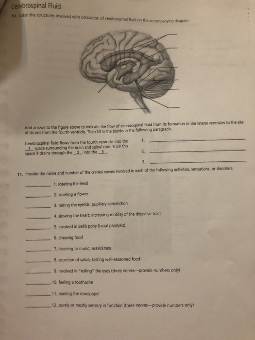
Label the structures involved with circulation of cerebrospinal fluid on the accompanying diagram Add arrows to the figure above to indicate the flow of cerebrospinal fluid from its formation in the lateral ventricles to the site of its exit from the fourth ventricle. Innermost vascular layer covering the brain.
Solved 290 Review Sheet 17 Ading Rubric Cerebrospinal Fluid Chegg Com
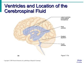
The main structures that make up the ventricular system include.
. Label the structures involved with circulation of cerebrospinal. 3 CSF flows through the paired lateral apertures or the single medial aperture and into the central canal of the spinal cord. Tough fibrous connective tissue 2.
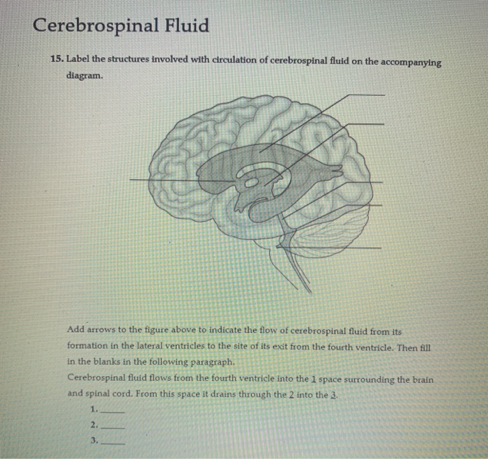
Identify the meningeal or associated structures described below. Place in order the circulation of cerebrospinal fluid. Add arrows to the figure above to indicate the flow of cerebrospinal fluid from its formation in the lateral ventricles to the site of its exit from the fourth ventricle.
Median foramen lateral foramen or central canal in any order g. 1109 Solutions 60 Chapters 108744 Studied ISBN. 14 Label the structures involved with circulation of cerebrospinal fluid on the accompanying diagram - Add arrows to the figure above to indicate the flow of cerebrospinal fluid from its formation in the lateral ventricles to the site of its exit from the fourth ventricle.
Then fill in the blanks in the following paragraph. Delicate and highly vascular 3. CSF enters ventricles.
Solution for Human Anatomy and Physiology Laboratory Manual Main Version 10th Edition Chapter 17 Problem 1. Structures instrumental in returning cerebrospinal fluid to the venous blood in the dural venous sinuses 4. Like a cobweb in structure arachnoid mater its outer layer forms the periosteum of the skull dura mater.
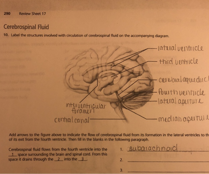
Most CSF enters subarachnoid space through the. Label the structures involved with circulation of cerebrospinal fluid on the accompanying diagram. Question not answered The correct answers are.
302 in Lab Manual Now label appropriately the structures involved with circulation of cerebrospinal. Follows every convolution 3. Composed of tough fibrous connective tissue 2.
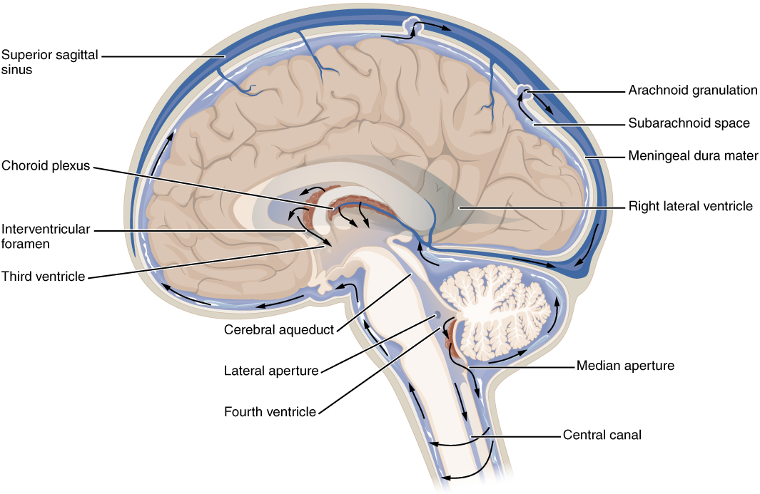
A dural fold that. Most CSF is secreted by the specialized tissue called the choroid plexus which is located within the lateral third and fourth ventricles. Tough fibrous connective tissue 2.
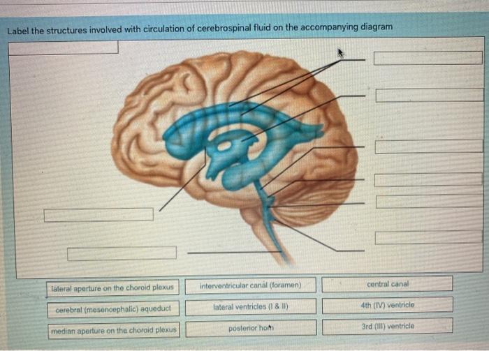
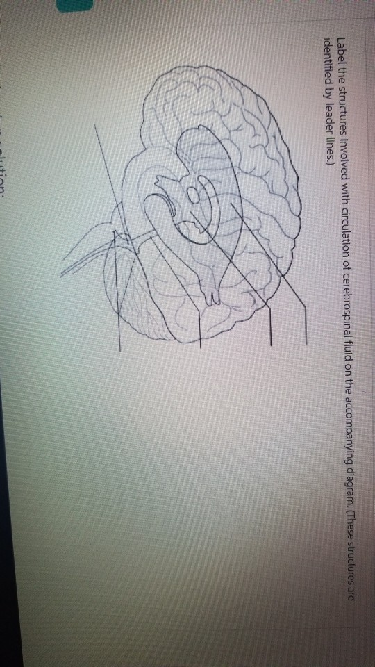
Ventricles subarachnoid space of the brain and spinal cord and the central canal of the spinal cord. Using the terms below correctly identify all structures indicated by leader lines on the diagram. These structures are identified by leader lines.
Delicate with cottony fibers 6. Chapter 17 Cerebrospinal_Fluid 1 Not my Question Bookmark. 9780321822338 Anatomy and physiology 5 1.
Arachnoid villus- drains cerebrospinal fluid into the venous blood in the dural venous sinuses 4. Like a cobweb in structure 6. Structures instrumental in returning cerebrospinal fluid to the venous blood in the dural sinuses arachnoid villi structure that forms the cerebrospinal fluid choroid plexus middle meninx.
It filters blood to form the plasma-like fluid that makes up CSF. Dura mater- outermost layer. Chapter 17 Cerebrospinal_Fluid 1 Not my Question Bookmark.
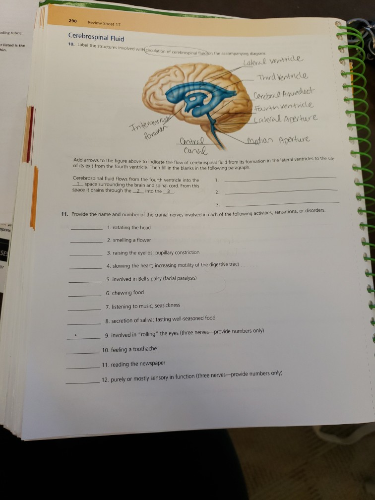
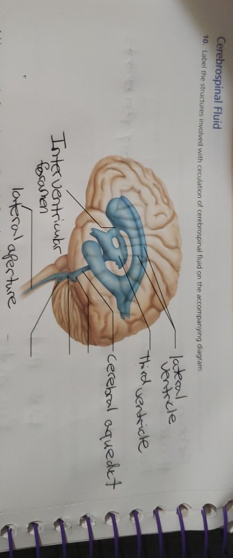
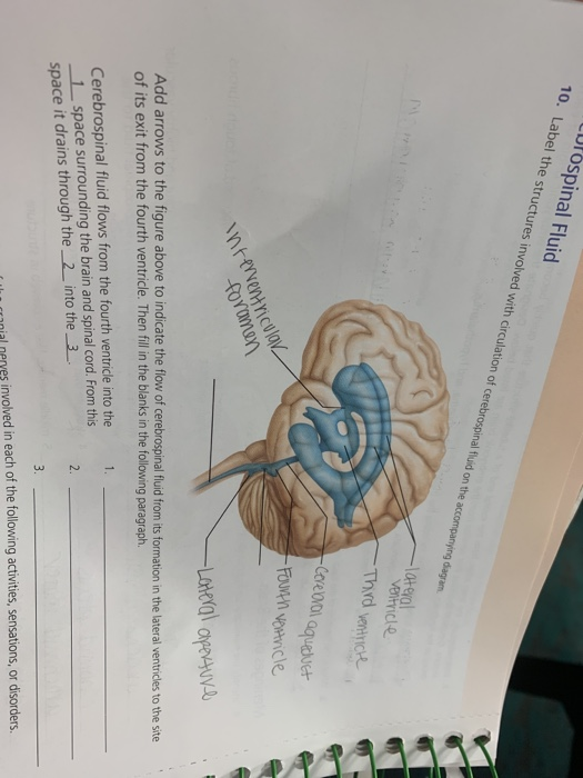
Label the structures involved with circulation of cerebrospinal fluid on the accompanying diagram lateconl Ventrcle Cerehsol oquedet resosmen afer ture. Pia mater- innermost vascular layer covering the brain. Structure that forms the cerebrospinal fluid 5.
Then fill in the blanks in the following paragraph. Structure that produces the cerebrospinal fluid 5. Structures that return cerebrospinal fluid to the venous blood.
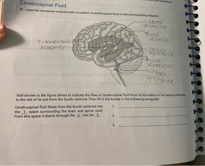
Add arrows to the figure above to indicate the flow of cerebrospinal fluid from its formation in the lateral ventricles to the site of its exit from the fourth ventricle. Outermost meninx covering the brain. From the subarachnoid space it drains through the ____ and into the ____.
Question 2 of 6 see Question 14 p. Label correctly the structures involved with circulation of cerebrospinal fluid on the accompanying diagram. The ventricular system is a network of communicating cavities within the brain called ventricles that function to produce transport and reabsorb cerebrospinal fluid CSF throughout the central nervous system.
These structures are identified by leader lines Question. Label the structures involved with circulation of cerebrospinal fluid on the accompanying diagram. Ventricular System Anatomy.
1114 Solutions 60 Chapters 98561 Studied ISBN. CSF passes through the arachnoid villi and is absorbed into the blood through the. Solution for Human Anatomy and Physiology Laboratory Manual Fetal Pig Version 11th Edition Chapter 17 Problem 1.
Then fillin the blanks in the following paragraph Cerebrospinal. Label the structures involved with circulation of cerebrospinal fluid on the accompanying diagram. Follows every convolution 3.
Plexus of each ventricle. CSF flows through subarachnoid space and baths outer surfaces of brain and spinal cord. Anatomy and Physiology questions and answers.
2 CSF flows from the third ventricle through the cerebral aqueduct into the fourth ventricle. See an animation of CSF circulation 38 Mb MOV movie Obs. Lateral and median apertures in walls of 4th ventricle.
CSF Circulation in light-green CSF lateral ventricles-- foramen of Monro third ventricle -- aqueduct of Sylvius -- fourth ventricle -- foramina of Magendie and Luschka -- subarachnoid space over brain and spinal cord -- reabsorption into venous sinus blood via arachnoid granulations. MT13 Clinical Anatomy and Physiology for Med Lab Science Laboratory Worksheet SU - ICLS Cerebrospinal Fluid 3. Drains cerebrospinal fluid into the venous blood in the dural venous sinuses 4.
A capillary bed that that lines the ventricles that secretes cerebrospinal fluid. Innermost meninx covering the brain. The cerebrospinal fluid flows from the 4th ventricle into the central canal of the spinal cord and the ____ space surrounding the brain and the spinal cord.
Identify the meningeal or associated structures described below. Anatomy and Physiology. Review_Exercise_14_pdf - Read File Online - Report Abuse.
Cerebrospinal fluid circulates through a system of cavities found within the brain and spinal cord. Label the structures involved with circulation of cerebrospinal fluid on the accompanying diagram. Some CSF enters the central canal of spinal cord.
1 CSF is produced by the choroid plexus in the ventricles. Label the structures involved with circulation of cerebrospinal fluid on the accompanying diagram. 9780321822321 Anatomy and physiology 5 1.
Solved Cerebrospinal Fluid 15 Label The Structures Involved Chegg Com
Solved Add Arrows To The Figureto Indicate The Flow Of Chegg Com
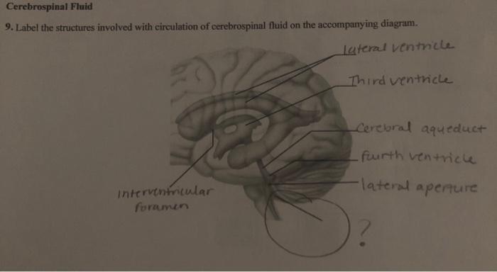
Solved Cerebrospinal Fluid 9 Label The Structures Involved Chegg Com

A P Lab Exercises 17 19 Flashcards Quizlet
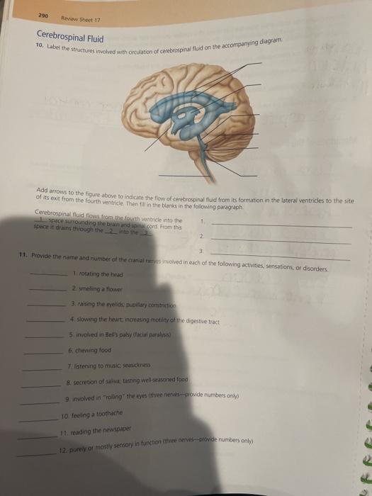
Solved 290 Review Sheet 17 Cerebrospinal Fluid 10 Label The Chegg Com
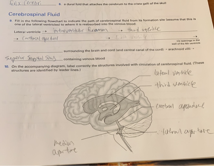
Solved Talx Lerebri 6 A Dural Fold That Attaches The Chegg Com
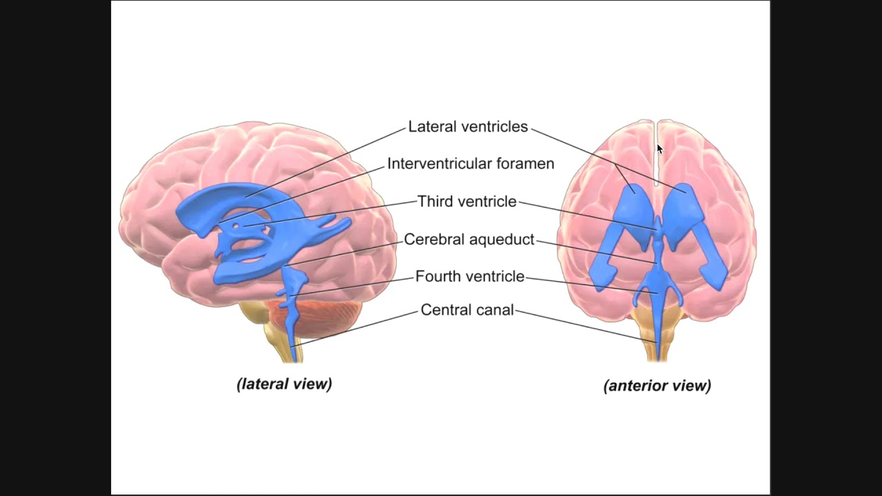
Circulation Of Cerebrospinal Fluid Csf Through Ventricles Youtube
Solved Cerebrospinal Fluid 10 Label The Structures Involved Chegg Com
Ppt On Cns
Solved Label The Structures Involved With Circulation Of Chegg Com
Solved Cerebrospinal Fluid 10 Label The Structures Involved Chegg Com
Solved Label The Structures Involved With Circulation Of Chegg Com
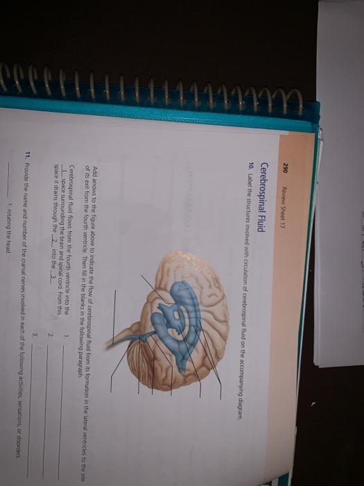
Solved 290 Review Sheet 17 Cerebrospinal Fluid 10 Label The Chegg Com
Solved Lutospinal Fluid 10 Label The Structures Involved Chegg Com
![]()
Cerebrospinal Fluid Flow Anatomy And Functions Kenhub

Cerebrospinal Fluid Flow An Overview Sciencedirect Topics

A P Lab Ex 17 Review Sheet Brain And Cranial Nerves Flashcards Quizlet
Circulation And Protection Of The Cns Anatomy And Physiology I
Solved The Cerebellum Cerebrospinal Fluid 15 Label The Chegg Com


Comments
Post a Comment